
Options in Medical Imaging - Enhance & Display In Specialized & High Resolution Applications
Image capture, processing, analysis, display, compression, and archival provide significant benefits to medical imaging. EPIX products offer all of these features to clinics and hospitals worldwide, in modalities such as: Nuclear Medicine (NM), Ultrasound (US), Computed Tomography (CT), Positron Emitted Tomography (PET), Magnetic Resonance Imaging (MR), and Radiography.
 |
|
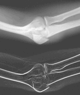 |
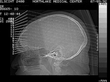
Top: X-Ray Computed Tomography image captured with SILICON VIDEO MUX. (Image courtesy of DataView.) Left Top: X-Ray image processed to enhance bone structure and minimize display of soft tissue. Left Bottom: X-Ray image processed to further emphasize bone structure. Images printed at 600 dpi with HP Laserjet 4 and EPIX software. |
For computed modalities, the imaging board acquires the image via a cable attached to the modality's monitor. This configuration does not affect the modality display. The imaging board acquires the image, provides processing capability, and displays on a secondary monitor.
Medical equipment manufacturers often use proprietary
video formats. Determining and setting parameters for proprietary
formats can be difficult; fortunately, EPIX engineers are experts
at nonstandard imaging and provide several tools to make the task
easier. Once a format is determined, it can be stored in a file
and later reloaded for immediate board setup.
Resolution & Pixel Clock Frequencies
EPIX imaging boards support thousands of image sizes:
128x128, 256x256, 512x512, 1024x1020, and others. All are selectable
through software control. Pixel clock frequencies between 12 MHz
and 30 MHz are appropriate for digitizing from most modalities;
both SILICON VIDEO MUX and 4MEG VIDEO span this range and more.
For higher resolution modalities, the SILICON VIDEO MUX accepts
pixel clock frequencies up to 40 MHz, supporting 1024x1020 resolution
at 30 frames per second. The 4MEG VIDEO Model 12 accepts pixel
clock frequencies to 50 MHz, providing even greater resolution.
Capture and display at 128x128, 256x256 or 512x512 resolution, are standard modes for NM, MR, US, PET, and CT imaging. With 4 megabytes of memory, studies of 256, 64, or 16 images, respectively, can be captured and stored as single files, each study being keyed to its particular patient. A 4MEG VIDEO Model 12, with 64 megabytes of image memory, provides four times this capacity! Radiography requires higher resolutions, such as 1024x1020, 2048x2048, or 4096x4096. Higher resolutions require extraordinary imaging boards, and the SILICON VIDEO MUX and 4MEG VIDEO are up to the task.
SVMUX Capture. Video Timing, & Display
Although the SILICON VIDEO MUX readily accepts nonstandard
video timing from the modality, it doesn't generate nonstandard
timing. Why is this important? Because horizontal and vertical
sync signals are required whenever an image is captured or displayed
- it's obvious that the SILICON VIDEO MUX must be connected to
the modality for image capture; it must also be connected to the
modality for any subsequent display (in order to access the modality's
nonstandard timing signals).
The SVMUX can capture and display images produced with pixel clock frequencies of 40 MHz or lower. Images with more lines than can be displayed on the monitor can be scrolled (moved up and down) to provide a view of the full resolution. An ideal configuration includes a monitor capable of displaying a full (nonwindowed) view.
4MEG VIDEO Capture, Video Timing, & Display
In contrast to the SILICON VIDEO MUX, the 4MEG VIDEO
can generate horizontal and vertical sync signals for nonstandard
video formats. As a result, the 4MEG VIDEO can duplicate the video
format generated by the modality, limited only by its maximum
pixel clock frequency of 50 MHz.
For example, assume an X-Ray film is scanned. A full 4096x4096 resolution is captured. The 4MEG VIDEO can provide display using one (or all four) of the following formats :
Option 1: High Resolution Capture with High Resolution Display
A 4096x4096 image can be captured and displayed at
2 frames per second. A 44 MHz pixel clock is required. There are
2 problems with this mode: a 4096x4096 monitor is a rare item,
and a display rate of 2 frames per second is too slow for comfortable
viewing.
Option 2: High Resolution Capture with Lower Resolution Display
For capture, the modality or camera provide video
timing and pixel clock; for display, the 4MEG VIDEO generates
timing and clock. This split capture / display approach to medical
imaging allows high resolution capture while providing comfortable
display. For our example, the 4096x4096 resolution is captured
at 2 frames per second while a 1300x976 display is provided at
30 frames per second. The 4MEG VIDEO provides both pan (the ability
to move the window left to right) and scroll (the ability to move
up and down) to provide visual access to the full image.
Option 3: High Resolution Capture With Standard RS-170 (CCIR)
Display
This mode combines high resolution capture with the
convenience and economy of standard display. A 640x480 (580) window
of the full 4096x4096 image is displayed on the monitor. The 4MEG
VIDEO's pan and scroll capability provides visual access to the
entire image.
Option 4: High Resolution Capture with Full Image Display
The 4MEG VIDEO can interpolate the high resolution
image into a lower resolution that can be displayed without pan
and scroll. Typical display resolutions are 1124x1124, 1024x1024,
580x580 (CCIR) and 480x480 (RS-170). The advantage of this display
is that you can see the full image; the disadvantage is that resolution
is sacrificed. If examination of the interpolated image reveals
a need for more detailed inspection, then Option 2 or 3 can always
be used. Capture resolution is not affected by interpolation,
so high resolution display is always an option.
Remote processing a Display
Teleradiology systems, which transmit images to remote
processing sites, need the 4MEG VIDEO in each receiving PC in
order to process, generate video timing for display, and to provide
image pan and scroll. If subsequent display is the only need,
processing is not required, and if the computer's VGA / Super
VGA monitor will be the display device, then an imaging board
is not necessary. Instead, a third party graphics package capable
of displaying TIFF images is all that is required.
Processing
EPIX imaging boards and software provide many functions
which enhance detail, reduce random noise, and provide accurate
measurement.
Histogram displays provide a graph of the distribution of captured grey levels. Several options for enhancing contrast are available. Random noise can be reduced by capturing a single image multiple times and averaging. Fourier Transforms, Inverse Fourier Transforms, and userprogrammable convolutions are provided. Calibrated distance and angle measurements can be taken directly from the computer image. Blob analysis functions provide reports on numbers, sizes, and positions of features.
Painting functions allow labeling of images and highlighting of significant areas for further analysis. Processing of selected areas of an image, as opposed to the complete image, are provided. Printing, at 600 dpi, is supported.
Other capabilities include gamma correction, background normalization, image subtraction, pseudocolor display, various filters and more. Images can be saved in a variety of formats including TIFF, X/Y, and ASCII. Lossless and lossy compression methods are available.
Summary
Subsequent processing is often desirable once a modality
has formed an image. Pixel by pixel corrections can be implemented
to provide better viewing. Features can be measured and the diagnoses
can be annotated. The image can be displayed, analyzed, printed,
compressed, transmitted over telephone lines, de-compressed, displayed,
processed, analyzed, compressed for storage, and stored in a digital
format.
EPIX products provide all of these capabilities (with the exception of the actual transmission over telephone lines). EPIX imaging boards and software provide high resolution image acquisition, exceptional image processing, and offer several alternatives for display - features valuable with all modalities.
EPIX Vision - April 1994 Newsletter
Specifications and prices subject to change without notice.
EPIX® imaging products are made in the USA.
Copyright © 2026 EPIX, Inc. All rights reserved.